The images are labeled providing an important medical and anatomical tool. Materials Microscope Slides LABPAQ Kit.

Heart Posterior View Diagram Quizlet
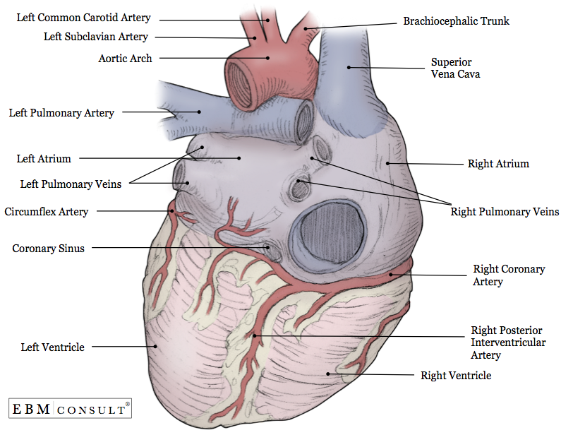
Anatomy of the human heart and coronaries.
. How to view anatomical structures. The regions of the body are labeled in boldface. The fat is light colored soft and without structure.
The regions of the body are labeled in boldface. It primarily functions as a lymphoid tissue although it has an important endocrine function that involves the production of thymosins. The human body is shown in anatomical position in an a anterior view and a b posterior view.
Using your forceps remove the fat that covers the upper part of the heart and blood vessels. Locate the external structures labeled on the anterior and posterior sides of the heart as shown below. This tool provides access to an MDCT atlas in the 4 usual planes allowing the user to interactively discover the heart anatomy.
Place the preserved heart on your dissecting tray. A body that is lying down is described as either prone or supine. The quiz mode makes it possible to evaluate the users progress.
The thymus is located anterior to the heart distal to the thyroid. We will view the histology during the lymphatic and immunity unit.
Anatomy Heart External
The Heart Chambers And Their Functions

Posterior View Of The External Heart Diagram Quizlet

4 Posterior View Of The Human Heart Download Scientific Diagram

Posterior View Of The Heart Heart Anatomy Heart Diagram Anatomy

Anatomy Of The Human Heart Posterior View Quiz

Heart Anatomy Anatomy And Physiology Ii

Posterior View Of The Heart Diagram Quizlet
0 komentar
Posting Komentar